A growing abdominal mass, an unexpected scan finding, or persistent pressure symptoms deserve more than a quick opinion about surgery. By the end, you will know which records and tests to expect, how specialists judge operability, and which questions help you compare treatment plans in Mumbai.
Key takeaways
- Request contrast-enhanced CT or MRI images, not only the written report.
- Confirm the biopsy plan with a sarcoma-focused multidisciplinary team.
- Ask which organs or blood vessels surgery may involve before consenting.
- Check experience with primary, recurrent and metastatic retroperitoneal sarcoma.
When an abdominal finding needs a sarcoma-focused assessment
A documented deep mass on ultrasound, CT or MRI, or measurable tumour growth on repeat imaging, warrants specialist review for retroperitoneal sarcoma assessment in Mumbai.
One symptom alone is less specific, but persistent abdominal or back pain, increasing abdominal fullness, early satiety, nausea, constipation, urinary symptoms, leg swelling or unexplained weight loss should not be ignored when they accompany an abnormal scan.
| Finding | What it means | Next step |
|---|---|---|
| Single nonspecific symptom | Many non-cancer conditions can cause it | Arrange routine medical assessment; escalate if it persists or worsens |
| Deep abdominal or retroperitoneal mass | The finding needs characterisation, even without symptoms | Request sarcoma-focused review before treatment |
| Measurable interval growth | Enlargement raises concern for an active tumour | Take repeat images and reports to a specialist promptly |
At the first consultation, the team will:
- Review previous scans, written reports and available pathology.
- Take a detailed symptom, cancer and family-history record.
- Examine your abdomen and assess relevant swelling or neurological findings.
- Check medicines, allergies and other medical conditions.
- Identify missing images, reports or biopsy records.
- Refer the case to a sarcoma-focused multidisciplinary team, not a surgeon alone.
That review may include a sarcoma surgeon, radiologist, pathologist, medical oncologist and radiation oncologist. Together, they assess the diagnosis, biopsy needs, resectability and alternatives before anyone recommends an operation.
Which scans and biopsy details shape the diagnosis
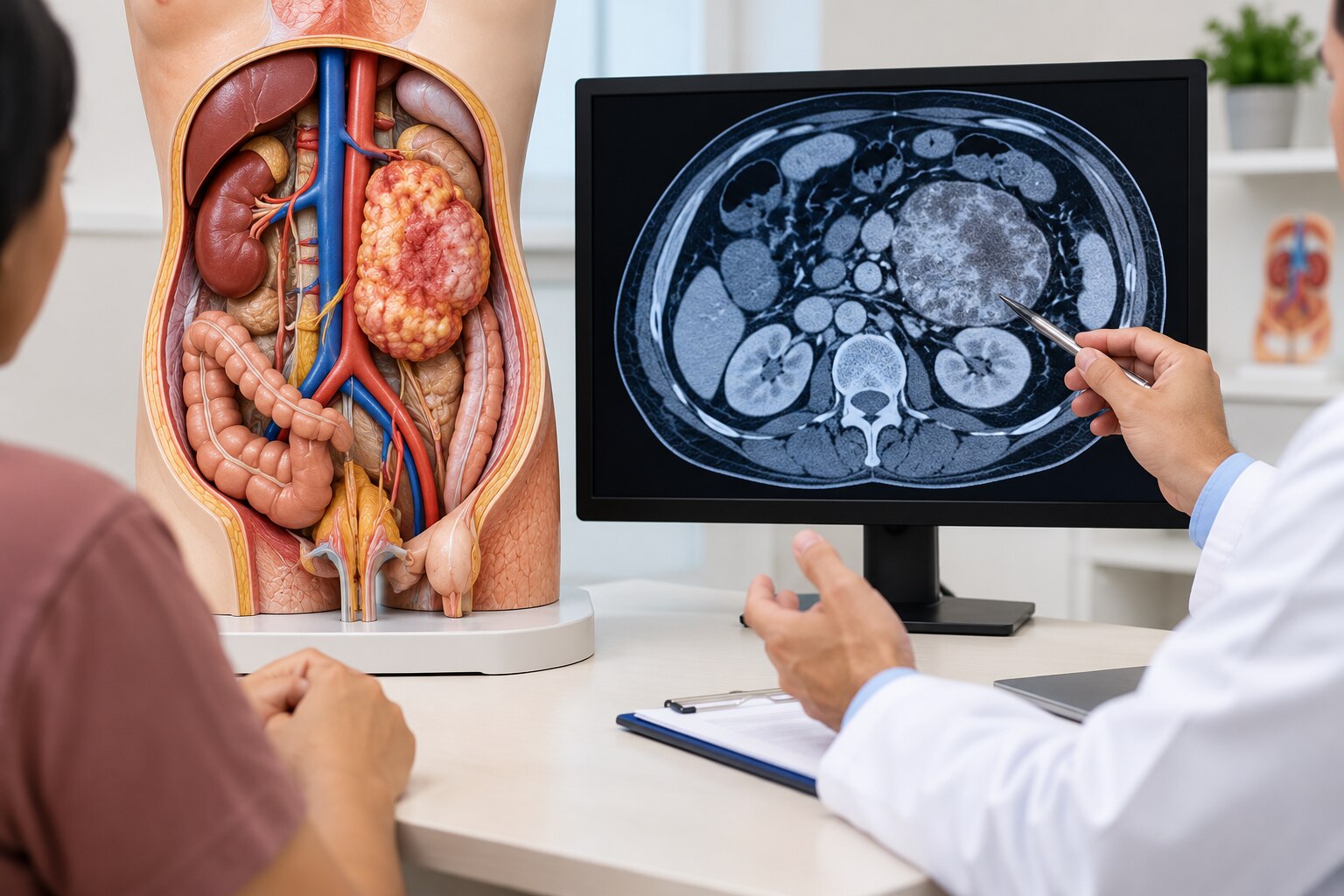
Contrast-enhanced CT of the abdomen and pelvis is usually the main planning study after the initial review. It maps the mass, nearby organs, blood vessels and tissue planes; chest CT or another appropriate chest image checks for lung spread.
| Study | Main purpose | Key limitation |
|---|---|---|
| Contrast-enhanced CT | Maps the tumour and abdominal relationships | May not show every soft-tissue plane clearly |
| Chest CT or suitable chest imaging | Looks for pulmonary metastases | Does not replace abdominal staging |
| MRI | Clarifies soft-tissue detail, tissue planes or a specific organ relationship | Adds selected information but does not replace high-quality CT for every tumour |
MRI becomes useful when CT leaves an important surgical question unanswered, such as whether a mass involves a particular organ or tissue plane. It is an additional tool, not an automatic substitute for CT.
A suspicious mass usually needs a planned, image-guided core-needle biopsy before definitive treatment. A core preserves tissue architecture for subtype and grade; fine-needle aspiration alone often provides too few cells.
The team selects a route that avoids major organs and vessels, and documents the entry site and track so the track can be removed with the tumour if surgery follows. An unplanned transperitoneal route can complicate the operation.
Pathology should name the sarcoma subtype and grade, report margin findings when tissue has already been removed, and test relevant molecular features. MDM2 amplification is especially important when well-differentiated or dedifferentiated liposarcoma is suspected. A retroperitoneal sarcoma treatment consultation in Mumbai should involve radiology, pathology and oncology specialists alongside the surgeon.
How the team decides whether surgery is realistic
A tumour is resectable when the team believes it can remove all visible disease safely, even if the operation requires nearby organs. Borderline resectable means complete removal might be possible but depends on surgical complexity, reconstruction and the patient’s expected recovery.
It is unsuitable for surgery when complete removal would cause unacceptable harm or disease is too widespread for meaningful local control.
Retroperitoneal sarcoma treatment planning looks beyond tumour size. The team assesses:
- Contact with or invasion of the kidney, ureter, pancreas, bowel, colon, major blood vessels, nerves and abdominal wall
- Sarcoma subtype and grade, which influence growth pattern and recurrence risk
- Multifocal disease or metastases, including spread to the lungs, liver or other sites
- Whether an operation can achieve complete macroscopic resection, meaning no visible tumour remains
The preferred operation removes the tumour intact, often en bloc with an organ directly invaded by it. Options can include kidney removal, vascular reconstruction, ureteric repair or reimplantation, and abdominal-wall reconstruction. Removing a kidney is not automatic; preserving it is not beneficial if it prevents safe, complete cancer removal.
Rupture or piecemeal removal can leave disease behind or seed the operative field.
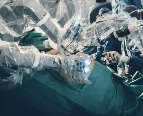
Laparoscopic or robotic surgery suits only selected small, favourably located tumours. Large tumours, vessel involvement or the need for an intact en-bloc specimen commonly require open surgery. The multidisciplinary team weighs cancer control against kidney function, bowel function, mobility and other lasting effects before recommending an operation.
How the plan changes for primary, recurrent or metastatic disease
A first operation for a primary retroperitoneal sarcoma is planned differently from treatment for recurrence or metastatic disease. The team must agree on the goal before choosing surgery, chemotherapy, radiation or observation.
1. Primary disease: Surgery may aim for complete macroscopic resection, often by removing the tumour intact with directly involved organs when that offers the best chance of local control. Kidney, bowel or vascular removal is not automatic; it depends on safe clearance and expected function.
2. Local recurrence: The team reviews previous operations, current imaging and the location of recurrent tumour. Repeat surgery may provide control when the disease is removable, but radiation, systemic treatment, observation or symptom control may be better when critical structures cannot be safely removed.
3. Metastatic disease: Lung, liver or other spread changes the balance between local treatment and whole-body treatment. Surgery for selected metastases can be discussed, but widespread or rapidly progressing disease often makes systemic therapy, symptom relief or observation the more realistic goal.
Chemotherapy is not automatically required before or after surgery. Its value depends on subtype, grade, stage, recurrence risk and drug sensitivity, separate from whether the tumour is technically removable.
Preoperative radiation is selective, not routine. The STRASS trial found no significant abdominal recurrence-free-survival benefit for the overall study population, so histology, anatomy, expected margins and treatment harm matter.
HIPEC or another intraperitoneal treatment is not a routine substitute for standard resection. Ask for evidence specific to your subtype and disease distribution.
What to verify before choosing a Mumbai specialist and proceeding
Before choosing a Mumbai specialist, ask for a written care plan rather than judging a clinic by location alone. A search for a retroperitoneal sarcoma specialist near Opera House should confirm who reviews your scans, biopsy and surgical options.
| Question to ask | What a clear answer includes |
|---|---|
| Is there a sarcoma multidisciplinary team? | Sarcoma surgeon, radiologist, sarcoma pathologist, medical oncologist and radiation oncologist review the case. |
| Are tests complete before surgery is proposed? | Contrast-enhanced CT of the abdomen and pelvis, chest imaging and a planned image-guided core-needle biopsy. |
| Who reviews the slides? | A sarcoma pathologist confirms subtype, grade and relevant molecular findings. |
| How will complex support be arranged? | Named vascular, urologic, colorectal, medical-oncology and radiation teams are available when needed. |
Ask whether the biopsy route and track were documented for removal with the specimen. An inadequately placed biopsy can complicate the operation.
Follow-up is part of retroperitoneal sarcoma care in Mumbai. Surveillance commonly uses CT of the abdomen and pelvis plus chest imaging every three to six months during the early years, then longer intervals when stable; subtype, grade, recurrence risk and treatment history determine the schedule.
A consultation with Cercano Surgical Oncology can organise specialist assessment, clarify resectability and separate the question “can surgery remove this safely?” from the assumption that surgery is automatically the answer.
Related service
Retroperitoneal sarcomas Comprehensive Care for Retroperitoneal Sarcomas Retroperitoneal sarcomas are rare cancers that develop in the retroperitoneum, the deep area of the... View service → |
Frequently asked questions
When does an abdominal finding need a sarcoma-focused assessment?
Arrange specialist review for a documented deep abdominal or retroperitoneal mass, measurable growth on repeat imaging, or a mass whose origin and relationship to nearby organs remain unclear.
Which scans and biopsy details matter for retroperitoneal sarcoma?
Bring the original CT or MRI images, contrast details and reports. Ask whether the biopsy uses image guidance, a planned needle path and tissue testing that can distinguish sarcoma subtypes.
How is surgical feasibility assessed?
The team reviews tumour size, location, involvement of the kidney, bowel, pancreas, major blood vessels and ureters, along with your fitness for surgery and the likelihood of complete tumour removal.
How does care differ for primary, recurrent or metastatic disease?
A first diagnosis focuses on staging and a safe treatment sequence. Recurrent disease requires comparison with prior operations and scans, while metastatic disease requires assessment of spread, symptoms and whether local or systemic treatment offers benefit.
What should you verify before choosing a Mumbai specialist?
Check the surgeon’s experience with retroperitoneal sarcoma, access to pathology and radiology review, multidisciplinary planning, hospital support for complex surgery, and a clear explanation of risks and alternatives.
Keywords